
Minimally Invasive Epidermal Cyst Removal in Hong Kong
Minimally Invasive Epidermal Cyst Removal in Hong Kong
2–3 mm Micro-Opening · Minimal Scarring · Approx. 20–30 Minutes
Have a persistent, growing lump under your skin that causes irritation or cosmetic concern?
An epidermal cyst (often colloquially called a “sebaceous cyst”) is a benign, keratin-filled sac beneath the skin. While medically harmless, cysts can enlarge, rupture, or become painfully infected if left untreated.
At Genesis Surgery, cyst removals are performed by a Specialist in General Surgery. Using a minimally invasive micro-excision technique through a 2–3 mm opening, we extract both the internal keratin contents and the entire cyst wall to minimise tissue trauma and post-procedural scarring.
✓ Specialist Surgeon: Performed by a Specialist in General Surgery
✓ Minimally Invasive: Small 2–3 mm micro-opening
✓ Procedure Duration: Approximately 20–30 minutes
✓ Anaesthesia: Local anaesthesia
✓ Recovery: Day procedure with immediate discharge
Recognising an Epidermal Cyst
Epidermal cysts commonly develop on the face, ears, scalp, neck, chest, and back. Typical features include:
- A smooth, firm, movable subcutaneous nodule
- A visible central dark pore or punctum (“blackhead”)
- Gradual enlargement over months or years
- Acute tenderness, redness, or purulent discharge if inflamed or infected
Important: Never squeeze or puncture a cyst at home. Forceful compression can rupture the fragile sac internally, releasing keratin into surrounding tissue and causing severe foreign-body inflammation, abscess formation, and noticeable scarring.
Differential Diagnosis: Is It an Epidermal Cyst?
Not every subcutaneous lump is an epidermal cyst. A clinical examination ensures accurate diagnosis and targeted treatment:
| Type of Lump | Key Distinguishing Characteristics |
|---|---|
| Epidermal Cyst | Firm, round, mobile nodule; often features a central punctum; filled with keratin. |
| Lipoma | Softer, lobulated, doughy mass located deeper in the subcutaneous fat layer. |
| Pilar Cyst | Firm, smooth nodule with a thicker wall, most frequently located on the scalp. |
| Dermatofibroma | Firm cutaneous nodule that characteristically dimples inward when pinched. |
Not sure whether your lump is an epidermal cyst?
A proper clinical assessment is important, as various subcutaneous lumps require different management approaches.
Enquire About a Clinical Assessment
Treatment: Minimally Invasive vs. Conventional Excision
Definitive treatment requires removing both the internal keratin contents and the entire surrounding cyst wall (capsule). Leaving any portion of the wall intact leads to high rates of recurrence.
| Conventional Excision | Minimally Invasive Technique | |
|---|---|---|
| Incision Size | Matches the diameter of the cyst | Approx. 2–3 mm |
| Cyst Wall Extraction | Excised intact through a wider wound | Dissected and delivered via micro-instruments |
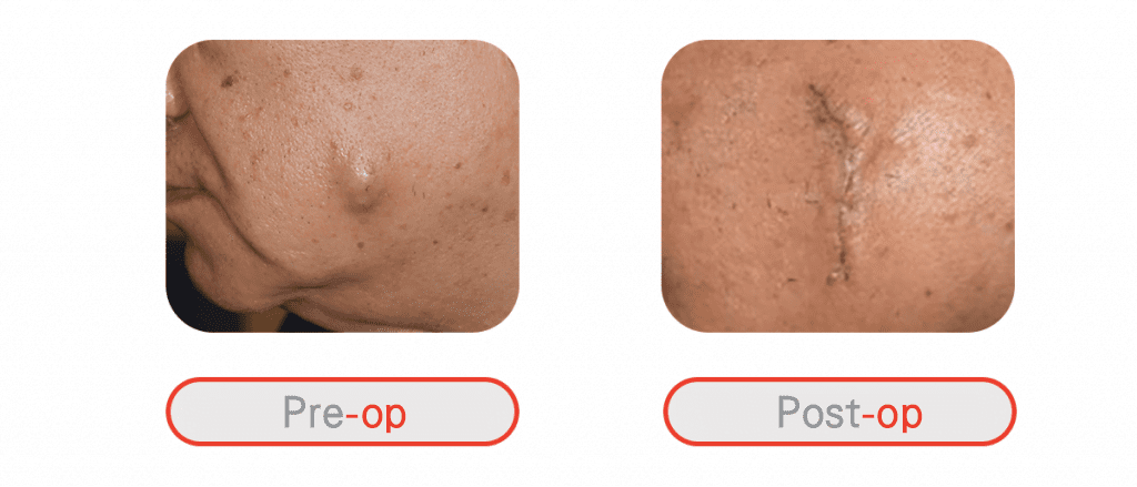
| Scar Profile | Noticeable linear scar | Minimal, discreet puncture mark |
| Recurrence Rate | Low when wall is fully excised | Low (~3% in published clinical data) |
| Downtime | Requires wound care for larger incision | Rapid healing; resume light routine next day |
Conventional Excision
Minimally Invasive Technique
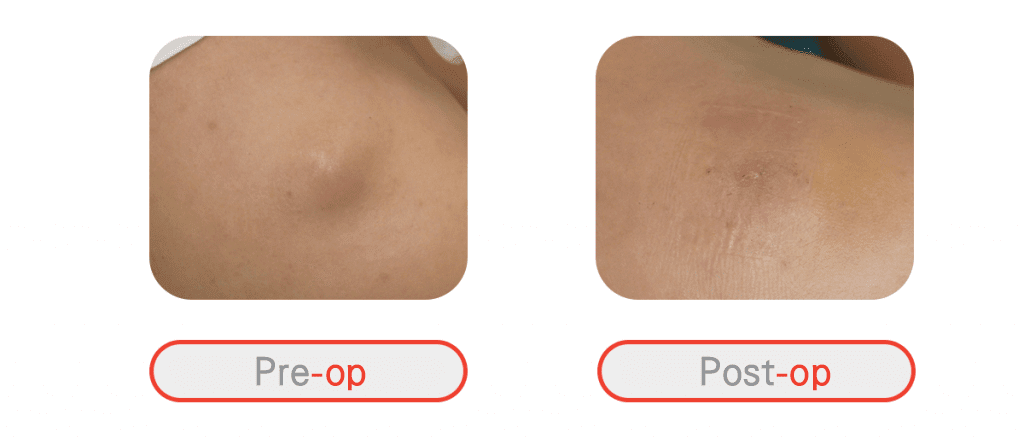
* The images are for reference only. Actual treatment outcomes depend on multiple factors, including cyst size, location, inflammation, wound healing and individual scar tendency.
What If My Epidermal Cyst Is Already Inflamed or Infected?
If a cyst is acutely infected or has formed an abscess, the capsule becomes friable and adherent to surrounding tissue, making immediate complete removal inadvisable. Treatment is carried out in two stages:
- Stage 1 (Acute Control): Minor incision and drainage (I&D) combined with clinical dressings and medication to eliminate infection and settle inflammation.
- Stage 2 (Definitive Removal): Once tissues have healed and softened (usually several weeks later), the remaining cyst wall is cleanly and permanently removed.
The Outpatient Procedure: Step-by-Step
- Clinical Assessment: The specialist surgeon examines the lump, reviews your medical history, and confirms suitability for micro-excision.
- Local Anaesthesia: Anaesthetic is administered locally around the lesion for a completely painless procedure.
- Micro-Excision (20–30 mins): A 2–3 mm opening is made over the apex. The keratin is decompressed, and the epithelial sac is gently freed and extracted intact.
- Wound Care & Same-Day Discharge: The micro-wound is dressed without bulky bandages. You may return home immediately.
- Follow-Up & Histology: Follow-up visits monitor wound healing. Where clinically indicated, excised tissue is submitted for histopathological testing to verify diagnosis.
Enquire About a Clinical Assessment
* All content on this website is provided for informational or educational purposes only. If you have questions about your condition or treatment, please consult a doctor or other qualified healthcare professional.
References:
- Choi MK, Chung KJ. Factors affecting complications after treatment of epidermal cyst. Yeungnam Univ J Med. 2019 May;36(2):136-140. doi: 10.12701/yujm.2019.00164. Epub 2019 Apr 1. PMID: 31620625; PMCID: PMC6784636. Available from: https://pubmed.ncbi.nlm.nih.gov/31620625/
- Hoang VT, Trinh CT, Nguyen CH, Chansomphou V, Chansomphou V, Tran TTT. Overview of epidermoid cyst. Eur J Radiol Open. 2019 Sep 5;6:291-301. doi: 10.1016/j.ejro.2019.08.003. PMID: 31516916; PMCID: PMC6732711. Available from: https://pubmed.ncbi.nlm.nih.gov/31516916/
- Wollina U, Langner D, Tchernev G, França K, Lotti T. Epidermoid Cysts – A Wide Spectrum of Clinical Presentation and Successful Treatment by Surgery: A Retrospective 10-Year Analysis and Literature Review. Open Access Maced J Med Sci. 2018 Jan 10;6(1):28-30. doi: 10.3889/oamjms.2018.027. PMID: 29483974; PMCID: PMC5816307. Available from: https://pmc.ncbi.nlm.nih.gov/articles/PMC5816307/
Why Choose Genesis Surgery?

Minimally Invasive Approach
For suitable cases, we use a minimally invasive technique involving an opening of approximately 2–3 mm, with the objective of completely removing the cyst while minimising unnecessary tissue damage and scarring.

Extensive Experience in Epidermal Cyst Removal
Genesis Surgery has accumulated extensive experience in removing epidermal cysts, with our medical team regularly engaging in professional and academic exchanges to introduce new management approaches.

Comprehensive Patient Care
We utilise approaches to reduce patient anxiety and discomfort during treatment, including music, VR goggles, vibration devices, and cooling devices.

Professional Follow-Up
To mitigate recurrence risks, we provide professional follow-up and maintain open communication regarding pre-treatment instructions, post-treatment wound care, and recovery monitoring.

Transparent Pricing
Treatment costs are explained clearly without hidden charges, allowing patients to understand expected costs before proceeding.
Frequently Asked Questions
Any procedure that punctures the skin can leave a mark. However, because minimally invasive removal uses a 2–3 mm micro-opening rather than a wide incision, tissue trauma is significantly reduced. The resulting mark is typically minimal and fades over time, depending on the cyst’s location and your natural healing tendency.